?

The Pulmonary Circulation and Selective Pulmonary Vasodilators


Dr. D. Blythe, FRACP, Senior Registrar

Dr. P.V. van Heerden, FFICANZCA, Staff Specialist Dept. of Intensive Care, Sir Charles Gairdner Hospital,
Hospital Ave., Nedlands, WA 6009
AUSTRALIA

Acknowledgement – This paper is based on a review article published by the authors in Anaesthesia and Intensive Care.


Introduction

Pulmonary hypertension (PHT) and hypoxaemia are common features of conditions such as the acute respiratory distress syndrome (ARDS) and persistent pulmonary hypertension of the newborn (PPHN).1,2 The use of selective pulmonary vasodilators (SPV) has recently advanced the therapeutic options available to the clinician dealing with conditions where PHT and hypoxaemia are factors.  

Pulmonary hypertension.

The normal pulmonary circulation is a low-resistance, high flow system which is able to accommodate passage of the cardiac output (CO) at a typical pressure of 20/12mmHg.3,4 Pulmonary hypertension (PHT) is defined as a mean pulmonary artery pressure (MPAP) greater than 20mmHg. PHT may be passive, secondary to increased pulmonary capillary pressure, which, in turn, may be due to elevated left atrial pressure. Alternatively, PHT may be an active phenomenon, in which case the primary pathological process resides at the precapillary or capillary level. Pulmonary arterial pressure normally increases with age and altitude. In a discussion of pulmonary hypertensive states, pulmonary vascular resistance (PVR) may be used as to describe the degree of obstruction to pulmonary blood flow. PVR is defined as

PVR = (Ppa - Pla ) / Q

where Ppa = pulmonary artery pressure, Pla = left atrial pressure, and Q = cardiac output. However, "resistance" is a concept derived from the fluid dynamics of laminar, non-pulsatile flow of a Newtonian liquid in channels with a constant radius. Blood is not a Newtonian fluid. Blood flow is pulsatile and turbulent, and the pulmonary vasculature does not have a constant radius. The use of PVR is therefore a simplification and subject to error, but justifiable as the errors become less significant as PVR rises, and the changes seen in pulmonary hypertensive states are of such magnitude that the assumptions made in determining PVR become less important. Nevertheless, estimates of PVR should always be interpreted in the light of supporting clinical information. In order for PHT to develop a number of homeostatic mechanisms in the pulmonary circulation have to be overcome. These mechanisms include -

In addition to these homeostatic mechanisms the pulmonary circulation is controlled by - When pulmonary arterial pressures are elevated by one or more of the above factors, the compensatory mechanisms of vessel distension and recruitment may be attenuated. Increased pulmonary blood flow may then result in PHT. In this setting the right ventricle (RV) may not be able to maintain flow against this elevated afterload and may fail. Most PHT is secondary to chronic pulmonary or cardiovascular disease. Primary PHT (PPH) is a disorder in which PAP is elevated in the absence of any demonstrable cause and accounts for a small proportion of cases. The more common causes of PHT are listed in Table 1.


Table 1. Causes of pulmonary hypertension


     I. Primary pulmonary hypertension

     II. Secondary pulmonary hypertension

      1. Parenchymal disease
        • Chronic airflow limitation
        • Fibrotic and granulomatous disorders
        • Upper airway obstruction
        • ARDS
      2. Vascular disease
        • Thromboembolus
        • Vasculitis
        • Congenital heart disease
        • Portal hypertension
        • Granulomatous pulmonary hypertension
        • Sickle cell disease
        • Toxin-induced disease
        • HIV infection
      3. Ventilatory disease
        • Kyphoscoliosis
        • Neuromuscular junction disorders
        • Restrictive pleural disorders
        • Sleep apnea
      4. Vascular obstruction
        • Mediastinal tumour
        • Aneurysms
        • Granulomata
        • Mediastinal Fibrosis
        • Pulmonary embolus
      5. Venous hypertension
        • Left ventricular failure
        • Pulmonary veno-occlusive disease
 

Pathophysiological mechanisms that contribute to the development of PHT are hypoxia via the mechanism of HPV, loss of, or mechanical distortion of vessels, or obstruction of vessels by thrombus.


Primary Pulmonary Hypertension (PPH)

PPH is a disorder where no aetiology for the PHT is evident. It occurs most commomly in women between the ages of 20 and 50. The condition is rare, prognosis is poor, and diagnosis is made by exclusion of other causes.7 The pathophysiological mechanism appears to be an abnormal proliferative response in the small muscular arteries in response to an unknown stimulus. Histological examination shows characteristic changes in both media and intima, as well as evidence of inflammation.8

Secondary Pulmonary Hypertension

  1. PHT in Cardiac Disease

    Cardiac disease causes PHT by both passive and active means. In passive PHT, such as in the case of pulmonary venous hypertension (the usual cause being left ventricular failure), longstanding venous hypertension requires an increased upstream pressure to maintain adequate pulmonary blood flow. In this setting there is pulmonary arterial intimal proliferation and vascular remodelling. Perivascular fibrosis occurs and sustained arterial hypertension results. Cardiac disease can also result in active PHT. Disorders characterised by left-to-right shunting such as septal defects, and patent ductus arteriosus also result in intimal thickening and fibrosis with smooth muscle hypertrophy. These features become irreversible, and flow becomes bidirectional or right-to-left. The resultant hypoxia compounds the PHT. Severe PHT with shunt reversal constitutes the Eisenmenger syndrome. It has also been suggested that chronic distension of vessels in the setting of left-to-right shunting may itself act as a stimulus to vasoconstriction. Acute increases in pulmonary blood flow are more likely to produce PHT than more gradual increases in flow due to the homeostatic mechanisms described above. Small vessel emboli may also contribute to the development of PHT.

  2. PHT in Pulmonary Thromboembolic Disease

    Pulmonary thromboembolism (PTE) is a common cause of PHT. The PTE syndromes can be classified according to which segment of the pulmonary vasculature is affected; small, intermediate or large arteries. The commonest occurrence is occlusion of pulmonary arteries of intermediate size by embolus. Such emboli generally originate from lower limb vessels and are often recurrent and multiple. PHT arises acutely, both from loss of cross-sectional area of the pulmonary vasculature, and from HPV secondary to the hypoxaemia caused by V/Q mismatch. Hypoxaemia is also contributed to by a number of other processes. Shunting is demonstrable in some patients and may occur from opening of a patent foramen ovale and intracardiac right-to left shunting as right atrial pressure rises due to pulmonary hypertension. Atelectasis from impaired surfactant production may cause areas of low V/Q ratio and contribute to shunting and V/Q mismatch. The fall in cardiac output which occurs with most pulmonary emboli can result in areas of high V/Q ratio and increased dead space which may also contribute to hypoxaemia. The role of humoral influences remains controversial. Thromboxane release from activated platelets at the site of embolism probably contributes directly to vasoconstriction and the development of PHT. The majority of survivors of massive, proximal vessel PTE survive because of clot resolution, in which case chronic PHT does not occur. 9

  3. PHT in Respiratory Failure

    There are several mechanisms by which respiratory disorders elicit PHT. For example, fibrotic or inflammatory disorders cause microvascular narrowing, leading ultimately to occlusion of vessels. Cross-sectional area available for flow is reduced, and, at the same time, remaining vessels are less distensible. In airflow limitation, V/Q mismatch causes hypoxaemia and reflex vasoconstriction. In asthma and emphysema, vessels are compressed by hyperexpanded lungs and in emphysema, destruction of parenchyma leads to concomitant loss of vessels. In hypoventilation syndromes, chronic hypoxia causes chronic pulmonary vasoconstriction, and eventually, fixed pulmonary hypertension.

    1. Hypoxic Pulmonary Vasoconstriction

      Alveolar hypoxia is a common feature of many respiratory disorders, and a major contributor to development of PHT. 10,11 Alveolar hypoxia occurs as a result of alveolar hypoventilation, ventilation/perfusion mismatch with increased physiological dead space or intrapulmonary shunting. Alveolar hypoxia produces a vasoconstrictor effect - so-called hypoxic pulmonary vasoconstriction (HPV). 12 HPV has the beneficial effect of diverting blood flow away from regions where the oxygen tension is low and thereby optimising V/Q matching. The vasoconstrictor response to hypoxia occurs mainly in small arterioles of about 200 µ m diameter.12 There are two major theories as to how alveolar hypoxia may bring about pulmonary vasoconstriction. Firstly, alveolar hypoxia may cause release of a vasoconstrictor substance. No such substance has been identified and there is now a reasonable amount of evidence that the response is mediated by decreased production of nitric oxide (NO). The second possibility is that hypoxia may stimulate cellular metabolism in vascular smooth muscle in such a way as to influence excitation-contraction coupling and directly cause vasoconstriction. The two mechanisms are not mutually exclusive. Other mechanisms also contribute to PHT in the chronic forms of respiratory failure such as increased blood viscosity due to polycythaemia, and PTE. Several of these mechanisms usually operate concurrently in chronic respiratory disease. HPV is often the major contributor in acute respiratory failure.  

    2. PHT and Acute Lung Injury

      Acute respiratory failure is commonly seen in the critically ill patient and results from a diffuse injury to lung parenchyma. The spectrum of disease that results is called acute lung injury (ALI), of which ARDS is the most severe form.13 The earliest abnormality of ALI is increased endothelial permeability, unless the precipitating cause is an injury to the alveolar epithelium. The neutrophil is suspected to be the initiator of this response, although it is worth noting that ARDS can occur in the neutropenic patient. It may be that tissue macrophages are also capable of initiating the process, and certain cytokines and endotoxin may be capable of causing membrane damage directly. Interstitial pulmonary oedema soon accumulates to the point where the lymphatics are overwhelmed and alveolar oedema occurs (the exudative phase). Early in the course of disease type I alveolar epithelial cells are reduced in number; parenchymal elasticity is reduced and the basement membrane is exposed. Lung compliance falls and atelectasis ensues. The accumulation of neutrophils in the pulmonary circulation is another early feature of the process. Surfactant abnormalities compound the process of alveolar collapse and flooding. Later, type II alveolar epithelial cells proliferate across the damaged basement membrane in an attempt at healing. Given an intact basement membrane and in the absence of ongoing insults, this would lead to regeneration of type I cells (the proliferative phase); in some patients, for unknown reasons, a dysfunctional response is seen which results in fibrosis. It is this sequence of events which causes the refractory hypoxaemia characteristic of ALI.14 However, the above are not the only mechanisms operating. At least some of the hypoxia In ALI is due to oedema-independent alterations in V/Q matching. Shunting of blood from injured vascular beds to relatively normal ones (HPV) is a critical protective mechanism in the normal or in the regionally injured lung. 12 HPV is particularly evident in the acute situation when metabolic acidosis and hypercarbia, both of which potentiate pulmonary vasoconstriction, are often present. In the diffusely-injured lung, the protective vasoconstrictor response can create an inappropriate widespread increase in vasomotor tone and increase pulmonary arterial pressures significantly. The stimulus for the enhanced HPV appears to be not only hypoxia, but also release of vasoactive mediators, such as platelet activating factor (PAF), tumour necrosis factor (TNF), interleukins 1,6 and 8 and thromboxane A2.15 Microvascular thrombosis is another feature of ALI, which may contribute to PHT.  

Management of PHT Associated with Acute Lung Injury

It has long been known that right ventricular (RV) systolic function is impaired in patients with PHT secondary to chronic lung disease, and, more recently, in patients with ALI. Impaired RV function has been demonstrated using both thermodilution techniques and radionuclide assessment. PHT is not usually severe in the patient with ALI, but some patients develop right ventricular failure and the prognosis is correlated with the degree of increase in PVR. Treatment of PHT in the patient with ALI is worthwhile, as lowering pulmonary arterial pressures (PAP) and the effective pulmonary capillary pressure improves RV function and possibly promotes resolution of pulmonary oedema.16,17 Until recently, the mainstays of treatment of PHT in ALI have been supportive, including maintenance of oxygenation and systemic vasodilator therapy. Phlebotomy as a therapeutic tool has fallen into disuse. Attempts at improving pulmonary circulation with cardiotonic agents (digoxin) and anticoagulation (warfarin) have not met with marked success. However, prophylactic anticoagulation has been advocated for patients with chronic PHT.

  1. Oxygen therapy

    Because hypoxia is a major contributor to both acute and chronic PHT, correction of hypoxia is an important component of management. However, patients with ALI gain only partial relief of hypoxia from delivery of a high fraction of inspired oxygen (FiO2). This is because the problem is one of intrapulmonary shunt and venous admixture.18 The specific effect of correcting hypoxia on haemodynamic performance is difficult to define in isolation. In early ALI, most of the increase in PVR can be abolished by perfusing the lungs with oxygenated blood19 , suggesting that HPV is responsible for the PHT. Later, when histological changes of fibrosis and vascular remodelling have occurred, the defect is not readily reversible. Accordingly, manipulation of haemodynamic and respiratory variables to achieve optimal oxygenation is crucial early in the management of ALI, before the oxygen transport problem becomes of sufficient duration to bring about fixed PHT.

  2. Systemic vasodilator therapy

    Although vasodilator therapy may lead to haemodynamic and symptomatic improvement in patients with chronic PHT, the effect is not universal, may not be sustained and there may be significant side-effects. Therapy must therefore be individualised and patient response monitored carefully. Various agents have been used as vasodilators in both primary and secondary PHT. These include -adrenergic antagonists, -adrenergic agonists, diazoxide, hydralazine, nitrates including NO, angiotensin-converting enzyme inhibitors, calcium channel blockers, prostaglandins, phentolamine, and adenosine. The ideal pulmonary vasodilator would decrease RV afterload while increasing CO and systemic oxygen delivery. To achieve these goals a substantial decrease in PAP would need to occur while stroke volume (SV) and systemic arterial pressure (SAP) remained unchanged. This combination of haemodynamic goals has proven difficult to achieve with systemically administered vasodilator agents. In patients with PPH undergoing right heart studies and vasodilator testing prior to long-term therapy, the "ideal" response is seen in only 25-30% of patients. However, when achieved, the "ideal" response is associated with regression of right heart abnormalities and improved survival.20-2 Although vasodilators can decrease PHT when administered systemically, their clinical usefulness is limited by their non-selectivity. There is a twofold requirement for selectivity. Agents should be selective for the pulmonary circulation (as opposed to the systemic). They should also be selective for well-ventilated alveoli as opposed to poorly-ventilated alveoli, thereby avoiding increased right-to-left shunt, venous admixture and worsening oxygenation. Several trials of intravenously infused agents (including agents infused directly into the pulmonary artery) have demonstrated that reductions in PAP can only be gained at the expense of systemic hypotension, with the concomitant risks of RV ischaemia and heart failure.23-5 Furthermore, increased flow to the areas of poor ventilation worsened V|Q mismatch. The use of systemic vasodilators in ALI has therefore largely been abandoned. An exception is adenosine. Adenosine is a synthetic purine nucleoside. Several studies describe its use as a pulmonary vasodilator in PPH.26,27

  3. Selective pulmonary vasodilators (SPV)

    1. Nitric oxide (NO)

      . Although currently not licensed for use in Australia, over 14,000 research papers have been written on NO in the last five years and it has become accepted for use as a SPV in ALI. NO is a gaseous, endothelium-derived smooth muscle relaxant which plays a key role in maintaining a basal level of systemic and pulmonary vascular relaxation as well as acting as an inflammatory mediator, a signalling molecule and a mediator of host defences. NO activates guanylate cyclase and stimulates production of cyclic GMP (c-GMP). In smooth muscle, c-GMP activates a protein kinase which reduces intracellular calcium concentrations, decreasing muscle contractility.28,29

      • Exogenous NO dilates the pulmonary vasculature when inhaled at low concentration.30,31 It is rapidly inactivated by binding to haemoglobin in the pulmonary circulation and is therefore devoid of systemic effect. Cardiac output and oxygen delivery are not increased, in contrast to intravenously administered prostacyclin. Also, because it is delivered to ventilated lung units, it has the effect of increasing blood flow in proportion to ventilation. The dual requirements for selectivity are thereby fulfilled. Of particular interest is the observation that PHT in ARDS is associated with impaired production of NO. The relevance of this observation is still uncertain, but it adds weight to the rationale for use of NO in ARDS.

      NO has other actions which include –

      • a bronchodilator effect, which has been demonstrated in animal studies and in humans with methacholine-induced bronchospasm, although the effect may not be as marked in man.
      • NO is a neurotransmitter in both central and peripheral nervous systems, and plays a role in macrophage toxicity.
      • regulation of leucocyte adhesion, platelet aggregation and smooth muscle proliferation.

      Inhaled NO has been shown to be an effective SPV in several clinical settings. The first report of its use to decrease PAP was in 1988, when Higgenbottam and co-workers reported that inhalation of NO decreased PAP in patients with PPH.32 The idea was subsequently developed using a number of experimental models.33 PHT was reversed without adverse systemic consequences in the majority of these models, confirming its potential as a SPV. Similar results have been obtained in humans. Rossaint et al.34 reported decreased PAP and improved oxygenation secondary to improved V/Q matching in ten patients with ALI. No adverse consequences were seen. Confirmation of Rossaint’s findings have been reported by Payen,35 and other groups. The focus has now shifted to establishing dose-response relationships and investigating potential toxicity. Of interest is the observation that NO results in improved PAO2 and reduced PA pressures, while cardiac output is unaffected. Intravenous prostacyclin, in contrast, has been shown to reduce PA pressures and increase oxygen delivery, principally by increasing cardiac output. The reasons for these differences in actions are not entirely clear. The initial work on NO has been done with doses ranging from 1 to 128 parts per million (ppm). More recently, using doses of between 60 and 230 parts per billion (ppb) Gerlach et al.36 demonstrated a significant improvement in oxygenation and decrease in shunt fraction without change in pulmonary resistance. Others have also noted that the doses of NO required to improve oxygenation are much lower than the doses required to reduce PAP. This suggests that the dose-response curve for pulmonary artery pressure may not be the same as that for oxygenation, and that the beneficial effect of NO on pulmonary perfusion is due to a redistribution effect, rather than pure enhancement of flow. In most cases, in adults, concentrations of 5-40ppm are sufficient to lower PAP. Concentrations as low as those used by Gerlach approach the level found in room air (usually around 10 ppb although levels in expired air can reach 250ppb). The response to NO is not entirely predictable. Several studies have now documented groups of critically ill patients with ALI who have not responded to low-dose NO (<40ppm).34,37 The mechanism of this variability in response to NO is not known, although the degree of initial PHT appears to correlate with the likelihood of response, those patients with the most marked PHT being most likely to benefit.37 Withdrawal of NO therapy has also proven to be problematic in patients with ALI and PHT. Sudden worsening of PHT and consequent hypoxaemia has been reported on cessation of therapy.37 This is an unpredictable phenomenon and the mechanism is not known. Possibilities include suppression of endogenous NO production by exogenous administration (product inhibition), or release of a vasoconstrictor substance. The toxicity of NO remains an area of active research. NO reacts with oxygen to form strong oxidants with as yet incompletely understood effects on normal and pathological cellular metabolism. The rate of oxidation is dependent on the oxygen concentration and the square of the NO concentration.91 At high concentrations, NO causes systemic methaemoglobinaemia and has caused death.38 At lower concentrations, the avid binding to haemoglobin may protect the systemic circulation from toxicity, but the pulmonary circulation and alveoli remain at risk. The clinical significance of reduced platelet adhesion following NO administration is not clear. 39 There are no reports of coagulation problems with NO in clinical use. Most toxicological studies have not addressed the issue of long-term NO administration, or the potential for accumulation over prolonged periods of therapy. There are studies reporting NO administration to infants for periods of up to several weeks, 40 and to adults with PPH in a pulsed fashion over periods of up to nine months, without adverse effect.41 However, the higher oxides of nitrogen are known to be toxic substances. Nitrogen dioxide (NO2) has considerable pulmonary toxicity, causing pneumonitis and pulmonary oedema, and has been reported as a cause of cytotoxicity and death in experimental animals.42.43 NO2 is metabolised to nitrous and nitric acids in aqueous solution, and a number of other higher nitrates and nitrites are formed with as yet unknown effects.44 Metabolism of NO to NO2 is therefore a potential hazard, and NO and NO2 monitoring are mandatory during use of NO, so that ambient levels of NO do not exceed 25ppm, nor NO2 levels exceed 1ppm. 45 Apart from potential toxicity, other problems with administration of NO include the delivery system required for its use and the high capital costs of delivering the agent. Although NO gas itself is currently not expensive, the cost of administration is significant.

    2. Prostacyclin

      Prostacyclin (PGI2) is a member of the prostaglandin family of lipid mediators derived from arachidonic acid. It is the main product of arachidonic acid metabolism in all vascular tissues studied so far. Endothelial cells produce most PGI2, but it is also synthesised by smooth muscle cells.46 PGI2 is the most potent known endogenous inhibitor of platelet.47-9 These effects are achieved by activation of intracellular adenylate cyclase, which is initiated when PGI2 binds to a specific cell-surface receptor. Adenylate cyclase catalyses production of cyclic AMP which activates a protein kinase, and decreases intracellular calcium levels. PGI2 is therefore a potent vasodilator in all vascular beds including the pulmonary circulation. The physiological role of prostacyclin is to maintain endothelial non-reactivity to platelets by inhibiting thrombus formation (while maintaining the capacity for vessel wall repair), and to maintain a low vascular resistance in beds such as the pulmonary circulation, where its secretion is increased by hypoxia.50 It also stimulates endothelial release of NO. Disorders of PGI2 production have been implicated in PHT although the significance of these observations remains uncertain. PGI2 has a short half-life (2-3 mins) and few side-effects. Catabolism of PGI2 begins with its spontaneous hydrolysis in plasma to 6-keto prostaglandin F1 (6-kPgF1). A number of oxidative steps follow, giving rise to a vasoconstrictor metabolite, 6-keto prostaglandin F2 (6-kPgF2 ). These reactions occur mainly in the liver and the inactive compounds are excreted in the urine. PGI2 metabolites can be measured using standard radioimmunoassay techniques (6-keto prostaglandin F1 , H-RIA-Kit, Advanced Magnetic, Cambridge, Mass, USA). PGI2 was first used intravenously as a pulmonary vasodilator in 1978.47 Used intravenously, it has been reported to be effective in animal models of PHT,51 and in humans with conditions including PPH,52 ARDS,17,18 heart failure,53 peripheral vascular disease, sepsis, and in patients after heart transplant. PGI2 has also been used intravenously to assess whether established PHT will respond to vasodilator therapy.44 In PPH, a beneficial effect has been shown in several trials.52 Trials in cardiac failure have failed to demonstrate reduced mortality,53 but there may be a place for the drug in the acutely decompensating patient in whom an agent with a short half-life, minimal toxicity and no tendency to develop tolerance is required. Similarly, PGI2 finds a role in pulmonary hypertensive crisis following cardiac surgery. 54 In the setting of ALI, Radermacher et al.16 demonstrated a decrease in PAP in patients with ARDS, associated with a fall in systemic pressure and a rise in CO. Overall, there was little net effect on oxygenation, as there was a marked deterioration in V/Q matching. The same group subsequently demonstrated improved RV function with PGI2 in a similar setting.17 Other investigators have had similar results.34 The lack of a beneficial effect on oxygenation when PGI2 is used intravenously and the emergence of NO as a selective agent prompted research into PGI2 administration via the inhaled route. The earliest report of the use of PGI2 via the inhaled route (inhaled aerosolised prostacyclin - IAP) appeared in 1978.48 A canine model in which administration of IAP led to a reversal of HPV and a decrease in PAP without systemic consequences was described in 1993.55 A case series of patients with ARDS reported favourable effects on MPAP and improved oxygenation during IAP therapy, again without systemic effect.56 Subsequent to this early work, animal trials have compared IAP with NO. 51,57 Shunt fraction was improved and a beneficial effect on oxygenation was noted for both agents. In humans, comparative trials in children58 and adults 59 with ARDS found that both NO and PGI2 acted as SPV’s, decreasing PAP without systemic effect and leading to improved oxygenation. A number of case reports have demonstrated the effectiveness of IAP for PHT and severe hypoxaemia in all age groups. 60-64 A clear dose-response relationship was defined in one patient.64 All reports so far are agreed that prostacyclin acts selectively and is at least as effective as NO. The main advantages of IAP over NO are ease of administration and lack of toxicity. Jet nebulisation, the mode of delivery for IAP, is a technique universally available and the equipment necessary for administration is minimal (Figure 1).

      Circuit for IAP administration

      Fig 1. Circuit for IAP Administration

      A = syringe pump with dilute prostacyclin solution; B = mechanical ventilator;
      C = jet nebuliser (continuously supplied with prostacyclin solution by pump A)
      D = gas supply for jet nebuliser.


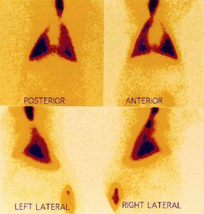
      The actual amount of drug deposited in the alveolus by such apparatus remains uncertain. A distal site of deposition is inferred from a knowledge of the particle size delivered and from the clinical effect observed. To achieve distal deposition a particle must be of size 0-5 µm.57 Even with appropriately-sized particles, the amount of a nebulised drug which actually reaches the alveolus in a ventilated patient is known to be a small fraction (less than 10%) of the total amount administered.65-7 The authors have performed nuclear medicine ventilation scans in piglets, using radiolabelled DTPA dissolved in the prostacyclin glycine buffer to determine site of drug deposition.68 A typical ventilation scan is shown in figure 2, indicating widespread and distal deposition of the tracer when delivered by jet nebuliser.

      V/Q scan showing jet nebuliser deposition

      Tracer Deposition with jet nebulisation
      (Technetium labelled DTPA in a piglet)

      As regards toxicity of IAP, systemic haemodynamic side-effects (hypotension, tachycardia) have not been seen with IAP. PGI2 is not transformed into an active form in the lung, nor is it significantly metabolised.69 Arterial concentrations of its major metabolite, 6-keto prostaglandin F1 have usually been reported as unchanged during inhalation of PGI2.55,70 Recent unpublished work from our institution describes arterial levels of 6-keto prostaglandin F1 in excess of 500 pg/ml during IAP therapy. These levels of 6-keto prostaglandin F1 have been shown to have a significant antiplatelet effect in vitro. The major area of concern has been the effect of IAP on coagulation.71 There has been no report of any clinically-important adverse effect on coagulation using PGI2 via the inhaled route. No other systemic toxic effects have been described. PGI2 has been reported as being irritant to the airway when administered intratracheally.72 A final area of concern is the alkalinity (pH 10.5) of the glycine buffer medium in which PGI2 is prepared. The authors have reported acute inflammation of the airways of piglets to which high dose IAP had been administered. 68

    3. Prostacyclin Analogues

      The emergence of PGI2 as a useful therapeutic agent has stimulated a research effort into design and synthesis of analogues with desirable physicochemical profiles. These include iloprost, cicaprost and beraprost.73-6 Iloprost is a stable carbacyclin derivative of PGI2. Like PGI2, it is found chiefly in vascular endothelium and has been shown to have similar effects on intracellular cAMP levels. Platelets and neutrophils exhibit in vitro responses to iloprost similar to PGI2, and the two agents are reported to have identical efficacy when used as pulmonary vasodilators. It has also been shown to be beneficial in peripheral vascular disease. Iloprost, being the more stable compound, has a longer duration of action when administered by the inhaled route. Beraprost has been tried orally in a small number of patients with PHT. It was successful in reducing PAP and improving cardiac index in a short-term study, and improved NYHA functional class in a long-term study. Minor side-effects were common but no significant haemodynamic disturbance was reported. Potential advantages of these PGI2 analogues over PGI2 include their solubility in saline, obviating the need for the highly alkaline buffer and the associated concerns regarding the toxicity of the buffer in which PGI2 is prepared, and a lower viscosity which facilitates nebulisation. None of these agents are commercially available for use in Australia. To date, only iloprost has been evaluated clinically to any extent. No cost analysis is available, and there are few controlled trials comparing the newer analogues with IAP or NO. Their clinical efficacy remains unknown, and they are regarded as investigational agents.

Conclusion

The advent of NO, and more recently IAP has provided clinicians, for the first time, with an opportunity to effectively treat the PHT and hypoxaemia characteristic of ALI, without major systemic adverse effect. Evidence now suggests that both NO and IAP act as selective pulmonary vasodilators, and their administration results in beneficial effects on haemodynamic performance and on oxygenation. The challenge for the future will be to define optimal use of these agents. For NO the main issues to be resolved are whether very low-dose NO is appropriate, and how best to prevent the toxic effects of the higher oxides. A simple delivery system is also needed. For IAP the main issues are defining a dose-response relationship, and ongoing documentation of its efficacy in the clinical setting, particularly compared with NO, the established agent. Other NO donors such as glyceryl trinitrate or sodium niroprusside may be just as effective via the inhaled route as NO, the current standard, and there is recent evidence in a rabbit model that both these agents are also successful selective pulmonary vasodilators.


References

  1. Zapol WM, Snider MT. Pulmonary hypertension in severe acute respiratory failure. N Engl J Med 1977: 296; 476-80.
  2. Zapol WM, Rie MA, Frikker M, Snider MT, Quinn DA. Pulmonary circulation during adult respiratory distress syndrome. In; Acute respiratory failure. Zapol WM, Falke KJ (eds.): Marcel Dekker 1985.
  3. Fishman AP. Pulmonary hypertension. In; The heart: Schlant RC, Alexander RW (eds), McGraw Hill, New York, 1994
  4. Nunn JF. The pulmonary circulation. In; Applied respiratory physiology: Butterworth Heinemann, Oxford, 1993.
  5. Benumof JL. Mechanism of decreased blood flow in atelectatic lung. J Appl Physiol 1988; 64: 68-77.
  6. West JB, Dollery CT. Distribution of blood flow and the pressure-flow relations of the whole lung. J Appl Physiol 1965; 20: 175-82.
  7. Rich S, Dantzker DR, Ayres SM et al. Primary pulmonary hypertension: A national prospective study. Ann Int Med 1987; 107: 216-23.
  8. Pietra GG, Edwards WD, Kay JM et al. Histopathology of primary pulmonary hypertension: A qualitative and quantitative study of pulmonary blood vessels from 58 persons in the National Heart, Lung and Blood Institute Primary Pulmonary Hypertension Registry. Circulation 1989; 80: 1198-206.
  9. Paraskos JA, Adelstein SJ, Smith RE et al. Late prognosis of acute pulmonary embolus. N Engl J Med 1973; 289: 55-58.
  10. Abraham AS, Cole RB, Green ID et al. Factors contributing to the reversible pulmonary hypertension in patients with acute respiratory failure studied by serial observation during recovery. Circ Res 1968; 23: 147-57.
  11. Fishman AP. Hypoxia on the pulmonary circulation - how and where it works. Circ Res 1976; 38: 221-31.
  12. Marshall BE, Marshall C, Frasch F, Hanson CW. Role of hypoxic pulmonary vasoconstriction in pulmonary gas exchange and blood flow distribution. Intens Care Med 1994; 20: 379-389.
  13. Bernard GR, Artigas A, Brigham KL et al. Report of the American-European consensus conference on ARDS: definitions, mechanisms, relevant outcomes and clinical trial coordination. Intens Care Med 1994: 20; 225-32.
  14. Kollef MH, Schuster DP. The acute respiratory distress syndrome. N Engl J Med 1995; 332: 27-37.
  15. Bernard GR, Reines HD, Halushka PV et al. Prostacyclin and thromboxane A2 formation is increased in human sepsis syndrome. Am Rev Resp Dis 1991; 144: 1095-101.
  16. Radermacher P, Santak B, Wust HJ, Tarnow J, Falke KJ. Prostacyclin for the treatment of pulmonary hypertension in adult respiratory distress syndrome: Effects on pulmonary capillary pressure and ventilation - perfusion distributions. Anesthesiology 1990; 72: 238-44.
  17. Radermacher P, Santak B et al. Prostacyclin and right ventricular function in patients with pulmonary hypertension associated with ARDS. Intens Care Med 1990; 16: 227-32.
  18. Dantzker DR, Brook CJ, Dehart P, Lynch JP, Weg JG. Ventilation-perfusion distributions in the adult respiratory distress syndrome. Am Rev Resp Dis 1979; 120: 1039-1052.
  19. Presberg K, Yanos J, Monk R, Corbridge T, Wood LDH. The role of mixed venous hypoxaemia in the pulmonary hypertension and shunt of canine low-pressure pulmonary oedema. Am Rev Resp Dis 1989; 139: A416.
  20. Weir EK, Rubin LJ, Ayres SM et al. The acute administration of vasodilators in primary pulmonary hypertension. Experience from the NIH registry on primary pulmonary hypertension. Am Rev Resp Dis 1989; 140: 1623-30.
  21. Rich S, Brundage BH. High-dose calcium channel blocking therapy for primary pulmonary hypertension: Evidence for long-term reduction in pulmonary arterial pressure and regression of right ventricular hypertrophy. Circulation 1987; 76: 135-41.
  22. Rich S, Kaufmann E, Levy PS. The effect of high doses of calcium channel blockers on survival in primary pulmonary hypertension. N Eng J Med 1992; 327: 76-81.
  23. Radermacher P, Huet Y, Pluskwa F. Comparison of ketanserin and sodium nitroprusside in patients with severe ARDS. Anesthesiology 1988; 68: 152-57.
  24. Radermacher P, Santak B, Becker H, Falke KJ. Prostaglandin E1 and nitroglycerin reduce pulmonary capillary pressure but worsen V/Q distributions in patients with ARDS. Anesthesiology 1989; 70: 601-606.
  25. Sibbald WJ, Driedger AA, McCallum D, Cunningham D, Cheung H. Nitroprusside infusion does not improve biventricular performance in patients with acute hypoxaemic respiratory failure. J Crit Care 1986;1: 197-203.
  26. Fullerton DA, Kirson LE, Jones SD, McIntyre RC. Adenosine is a selective pulmonary vasodilator in cardiac surgical patients. Chest 1996; 109: 41-46.
  27. Nootens M, Schrader B, Kaufmann E, Vestal R, Long W, Rich S. Comparative acute effects of adenosine and prostacyclin in primary pulmonary hypertension. Chest 1995; 107: 54-57.
  28. Moncada S, Palmer RMJ, Higgs EA. Nitric oxide: Physiology, pathophysiology, and pharmacology. Pharmacological Reviews 1991; 43: 109-42.
  29. Kam PCA, Govender G. Nitric oxide: basic science and clinical applications. Anaesthesia 1994; 49: 515-21.
  30. Fink MP, Payen D. The role of nitric oxide in sepsis and ARDS: synopsis of a roundtable conference held in Brussels on 18-20 March 1995. Intens Care Med 1996; 22: 158-65.
  31. Fratacci MD, Frostell CG, Chen TY, Wain JC, Robinson DR, Zapol WM. Inhaled nitric oxide. A selective pulmonary vasodilator of heparin-protamine vasoconstriction in sheep. Anesthesiology 1991; 75: 990-99.
  32. Higenbottam TW, Pepke-Zaba J, Scott J, Woolman P, Coutts C, Wallwork J. Inhaled endothelium-derived relaxing factor in primary pulmonary hypertension. Am Rev Resp Dis 1988; 137: A107.
  33. Frostell C, Fratacci MD, Wain JC, Jones R, Zapol WM. Inhaled nitric oxide; a selective pulmonary vasodilator reversing hypoxic pulmonary vasoconstriction. Circulation 1991; 83: 2038-47.
  34. Rossaint R, Falke K, Lopez F, Slama K, Pison U, . Inhaled nitric oxide for the adult respiratory distress syndrome. N Eng J Med 1993; 328: 399-405.
  35. Payen D, Gatecel C, Guinard N. Inhalation of low dose of nitric oxide and IV L-arg in ARDS patients. Am Rev Resp Dis 1993; 147: A720.
  36. Gerlach H, Pappert D, Lewandowski K, Rossaint R, Falke KJ. Long-term inhalation with evaluated low doses of nitric oxide for selective improvement of oxygenation in patients with adult respiratory distress syndrome. Intens Care Med 1993; 19: 443-49.
  37. Bigatello LM, Hurford WE, Kacmarek RM, Roberts JD, Zapol WM. Prolonged inhalation of nitric oxide in patients with severe adult respiratory distress syndrome. Anesthesiology 1994; 80: 761-70.
  38. Clutton-Brock J. Two cases of poisoning by contamination of nitrous oxide with the higher oxides of nitrogen during anaesthesia. Br J Anaesth 1967; 39: 388-92.
  39. Samama CM, Diaby M, Fellahi JL et al. Inhibition of platelet aggregation by inhaled nitric oxide in patients with ARDS. Anesthesiology 1995; 83: 56-65.
  40. Abman SH ,Griebel JL, Parker DK, Schmidt JM, Swanton D, Kinsella JP. Acute effects of inhaled nitric oxide in children with severe hypoxaemic respiratory failure. J Pediatr 1994; 124: 881-88.
  41. Channick RN, Newhart JW, Johnson F et al. Pulsed delivery of inhaled nitric oxide in patients with primary pulmonary hypertension. Chest 1996; 109: 1545-49.
  42. Shiel FOM. Morbid anatomical changes in the lungs of dogs after inhalation of higher oxides of nitrogen during anaesthesia. Br J Anaesth 1967; 39: 413-24.
  43. Greenbaum R, Bay J, Hargreaves MD et al. Effects of higher oxides of nitrogen on the anaesthetised dog. Br J Anaesth 1967; 39: 393-403.
  44. Gaston B, Drazen JM, Loscalzo J, Stamler JS. The biology of nitrogen oxides in the airway. Am J Resp Crit Care Med 1994; 149: 538-51.
  45. Centres for Disease Control. Recommendations for occupational safety and health standards MMWR 1988;37(suppl):21.
  46. Vane JR, Botting RM. Pharmacodynamic profile of prostacyclin. Am J Cardiol 1995; 75: 3A-10A.
  47. Szczeklik A, Gryglewski RJ, Nizankowska E, Nizankowski R, Musial J. Pulmonary and anti-platelet effects of intravenous and inhaled prostacyclin in man. Prostaglandins 1978; 16: 651-60.
  48. Szczeklik A, Gryglewski RJ, Nizankowski R, Musial J, Pielan R, Mruk J. Circulatory and anti-platelet effects of intravenous prostacyclin in healthy men. Pharm Res Comm 1978; 10: 545-56.
  49. Tremoli E, Colli S, Paoletti R. Mode of action of PGI2 and its stable derivative iloprost on platelets and leukocytes. Thrombosis Research 1990; Suppl XI: 33-42.
  50. Hyman AL, Spannhake EW, Kadowitz PJ. Prostaglandins and the lung. Am Rev Resp Dis 1978; 117: 111-36.
  51. Zobel G, Dacar D, Rodl S, Friehs I. Inhaled nitric oxide versus inhaled prostacyclin and intravenous versus inhaled prostacyclin in acute respiratory failure with pulmonary hypertension in piglets. Pediatric Research 1995; 38: 198-204.
  52. Long W, Rubin LJ et al. Randomized trial of conventional therapy alone vs. conventional therapy and continuous infusions of prostacyclin in primary pulmonary hypertension . Am Rev Resp Dis 1993; 147: A538.
  53. Haywood GA, Adams KF, Gheorghiade M, McKenna WJ. Is there a role for epoprostenol in the management of heart failure ? Am J Cardiol 1995; 75: 44A-50A.
  54. Goldman AP, Delius RE, Deanfield JE, Macrae DJ. Nitric oxide is superior to prostacyclin for pulmonary hypertension after cardiac operations. Ann Thor Surg 1995; 60: 300-306.
  55. Welte M, Zwissler B, Habazettl H, Messmer K. PGI2 aerosol versus nitric oxide for selective pulmonary vasodilation in hypoxic pulmonary vasoconstriction. Eur Surg Res 1993; 25: 329-39.
  56. Walmrath D, Schneider T, Pilch J, Grimminger F, Seeger W. Aerosolised prostacyclin in adult respiratory distress syndrome. Lancet 1993; 342: 961-62.
  57. Zwissler B, Welte M, Messmer K. Effects of inhaled prostacyclin as compared with inhaled nitric oxide on right ventricular performance in hypoxic pulmonary vasoconstriction. J Cardiothorac and Vascular Anaesth 1995; 9: 283-89.
  58. Pappert D, Busch T, Gerlach H, Lewandowski K, Radermacher P, Rossaint R. Aerosolized prostacyclin versus inhaled nitric oxide in children with severe acute respiratory distress syndrome. Anesthesiology 1995; 82: 1507-11.
  59. van Heerden PV, Blythe D, Webb SAR. Inhaled aerosolized prostacyclin and nitric oxide as selective pulmonary vasodilators in ARDS - a pilot study. Anaesth Intens Care 1996; 24: 564-68.
  60. Bindl L, Fahnenstich H, Peukert U. Aerosolised prostacyclin for pulmonary hypertension in neonates. Arch Dis Childhood 1994; 71: F214-16.
  61. Webb SAR, Stott S, PV van Heerden. The use of inhaled aerosolized prostacyclin (IAP) in the treatment of pulmonary hypertension secondary to pulmonary embolism. Intens Care Med 1996; 22: 353-55.
  62. Walmrath D, Schneider T, Pilch J, Schermuly R, Grimminger F, Seeger W. Effects of aerosolized prostacyclin in severe pneumonia. Am J Resp Crit Care Med 1995; 151: 724-31.
  63. Bein T, Keyl C, Pfeiefer M, Metz C. Administration of aerosolized prostacyclin in patients with severe respiratory failure and pulmonary hypertension. Chest 1995; 108: 119S.
  64. van Heerden PV, Webb SAR, Hee G, Corkeron M, Thompson WR. Inhaled aerosolized prostacyclin as a selective pulmonary vasodilator for the treatment of severe hypoxaemia. Anaesth Intens Care 1996; 24: 87-90.
  65. Coleman DM, Kelly HW, McWilliams BC. Determinants of aerosolized albuterol delivery to mechanically ventilated infants. Chest 1996; 109(6): 1607-13.
  66. Fuller HD Dolovich MB, Posmituck G, Pack WW, Newhouse MT. Pressurised aerosol versus jet aerosol delivery to mechanically ventilated patients - comparison of doses to the lungs. Am Rev Resp Dis 1990; 141: 440-44.
  67. Watterberg KL, Clark AR, Kelly HW, Murphy S. Delivery of aerosolized medication to intubated babies. Ped Pulmonol 1991; 10: 136-41.
  68. Piglet study
  69. Bahkle YS Ferreira SH. Lung metabolism of eicosanoids. In; Fishman AP, Fisher AB (eds): Handbook of physiology, section 3, Am Phys Soc, Bethesda, 1985.
  70. Habler O, Kleen M, Zwissler B et al. Inhalation of prostacyclin (PGI2) for 8 hours does not produce signs of acute pulmonary toxicity in healthy lambs. Intens Care Med 996; 22: 426-433.
  71. Antiplatelet effect
  72. Gardiner PJ, Copas JL, Elliott RD, Collier HOJ. Tracheobronchial irritancy of inhaled prostaglandins in the conscious cat. Prostaglandins 1978; 15: 303-15.
  73. Olschewski H, Walmrath D, Schermuly R, Ghofrani A, Grimminger F, Seeger W. Aerosolized prostacyclin and iloprost in severe pulmonary hypertension. Ann Int Med 1996; 124: 820-24.
  74. Hildebrand M, Nieuweboer B, Schutt A. Development and validation of a sensitive and specific radioimmunoassay for the determination of cicaprost in biological samples. J Immunoassay 1994; 15: 171-90.
  75. Drews G, Spiegel HU, Hermsdorf T, Dettmer D, Richter V, Hauss J. Cytoprotective effects of a stable prostacyclin analog and a calcium channel blocker: A study on isolated rat hepatocytes. Transplantation Proceedings 1995; 27: 2799.
  76. Saji T, Ozawa Y, Nakayama T et al. Short and long-term effects of the new oral prostacyclin analogue, beraprost sodium, in patients with severe pulmonary hypertension. J Cardiol 1996; 27: 197-205.